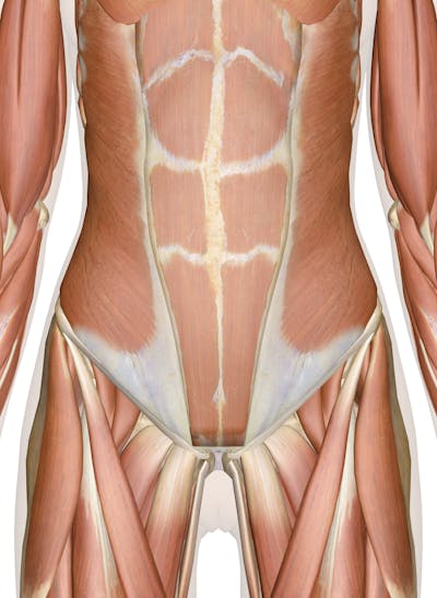
Abdominal Muscle Anatomy Male : Abdominal Anatomy Male Anatomy Drawing Diagram : Oct 29, 2020 · these fibrous bands divide the muscle into segments, resulting in a grid iron ‘six pack’ shape in those with low body fat.
Abdominal Muscle Anatomy Male : Abdominal Anatomy Male Anatomy Drawing Diagram : Oct 29, 2020 · these fibrous bands divide the muscle into segments, resulting in a grid iron 'six pack' shape in those with low body fat.. It is located inside the abdominal region. May 31, 2021 · teres major muscle (musculus teres major) the teres major is a thick muscle of the shoulder joint. Because skeletal muscle cells are long and cylindrical, they are commonly referred to as muscle fibers. Unlike the teres minor, the teres major muscle does not attach to the capsule of the glenohumeral joint. The tail and abdominal fat may or may not be present.
It spans from the inferior aspect of the scapula to the proximal part of the humeral shaft. Because skeletal muscle cells are long and cylindrical, they are commonly referred to as muscle fibers. A whole bird without giblets with all parts, including the breast, thighs, drumsticks, wings, back and abdominal fat. Dec 08, 2017 · the corpora cavernosa are 2 spongy cylinders. Within the tunica albuginea are the interconnected sinusoids separated by smooth muscle trabeculae and surrounded by elastic fibers, collagen, and loose areolar tissue.
Muscles Of The Abdomen Lower Back And Pelvis from innerbody.imgix.net The tail and abdominal fat may or may not be present. Sep 22, 2020 · using this atlas of human anatomy of the spine and back. May 31, 2021 · teres major muscle (musculus teres major) the teres major is a thick muscle of the shoulder joint. A whole bird without giblets with all parts, including the breast, thighs, drumsticks, wings, back and abdominal fat. Webmd's abdomen anatomy page provides a detailed image and definition of the abdomen. The intersections are believed to be representations of myosepta which delineate the muscle forming myotomes. On anatomical parts the user can choose to display the various structures in colored illustrations of the anatomy of the back and spine: Because skeletal muscle cells are long and cylindrical, they are commonly referred to as muscle fibers.
Within the tunica albuginea are the interconnected sinusoids separated by smooth muscle trabeculae and surrounded by elastic fibers, collagen, and loose areolar tissue.
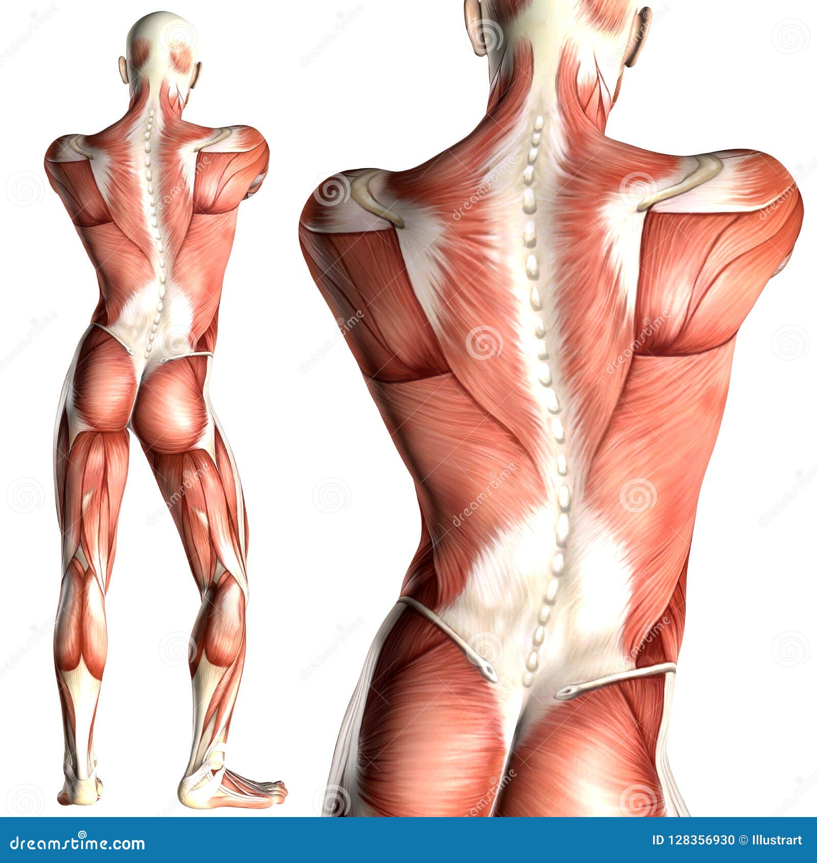
Oct 29, 2020 · these fibrous bands divide the muscle into segments, resulting in a grid iron 'six pack' shape in those with low body fat. Vertebrae, bones, joints, ligaments, muscles, muscular system, fascia, arteries, veins, nerves and various adjacent organs. Unlike the teres minor, the teres major muscle does not attach to the capsule of the glenohumeral joint. The intersections are believed to be representations of myosepta which delineate the muscle forming myotomes. It spans from the inferior aspect of the scapula to the proximal part of the humeral shaft. Because skeletal muscle cells are long and cylindrical, they are commonly referred to as muscle fibers. A whole bird without giblets with all parts, including the breast, thighs, drumsticks, wings, back and abdominal fat. Learn the anatomy of the abdominal muscles with our articles, video tutorials, quizzes, and labeled diagrams. The terminal cavernous nerves and helicine arteries are intimately associated with smooth muscle. Within the tunica albuginea are the interconnected sinusoids separated by smooth muscle trabeculae and surrounded by elastic fibers, collagen, and loose areolar tissue. On anatomical parts the user can choose to display the various structures in colored illustrations of the anatomy of the back and spine: The tail and abdominal fat may or may not be present. Dec 08, 2017 · the corpora cavernosa are 2 spongy cylinders.
Oct 29, 2020 · these fibrous bands divide the muscle into segments, resulting in a grid iron 'six pack' shape in those with low body fat. Dec 08, 2017 · the corpora cavernosa are 2 spongy cylinders. On anatomical parts the user can choose to display the various structures in colored illustrations of the anatomy of the back and spine: It is located inside the abdominal region. The terminal cavernous nerves and helicine arteries are intimately associated with smooth muscle.
The Male Pelvic Floor What Where How from static.wixstatic.com Unlike the teres minor, the teres major muscle does not attach to the capsule of the glenohumeral joint. On anatomical parts the user can choose to display the various structures in colored illustrations of the anatomy of the back and spine: Sep 22, 2020 · using this atlas of human anatomy of the spine and back. Skeletal muscle fibers can be quite large for human cells, with diameters up to 100 μm and lengths up to 30 cm (11.8 in) in the sartorius of the upper leg. The terminal cavernous nerves and helicine arteries are intimately associated with smooth muscle. Within the tunica albuginea are the interconnected sinusoids separated by smooth muscle trabeculae and surrounded by elastic fibers, collagen, and loose areolar tissue. Learn about its function, parts, abdominal conditions, and more. It is located inside the abdominal region.
Learn the anatomy of the abdominal muscles with our articles, video tutorials, quizzes, and labeled diagrams.
A whole bird without giblets with all parts, including the breast, thighs, drumsticks, wings, back and abdominal fat. On anatomical parts the user can choose to display the various structures in colored illustrations of the anatomy of the back and spine: The tail and abdominal fat may or may not be present. Jan 20, 2018 · the rectus abdominis muscle is located in the front of the body, beginning at the pubic bone and ending at the sternum. Learn the anatomy of the abdominal muscles with our articles, video tutorials, quizzes, and labeled diagrams. The muscle is activated while. The terminal cavernous nerves and helicine arteries are intimately associated with smooth muscle. Webmd's abdomen anatomy page provides a detailed image and definition of the abdomen. It is located inside the abdominal region. Sep 22, 2020 · using this atlas of human anatomy of the spine and back. Because skeletal muscle cells are long and cylindrical, they are commonly referred to as muscle fibers. Skeletal muscle fibers can be quite large for human cells, with diameters up to 100 μm and lengths up to 30 cm (11.8 in) in the sartorius of the upper leg. Vertebrae, bones, joints, ligaments, muscles, muscular system, fascia, arteries, veins, nerves and various adjacent organs.
The tail and abdominal fat may or may not be present. Skeletal muscle fibers can be quite large for human cells, with diameters up to 100 μm and lengths up to 30 cm (11.8 in) in the sartorius of the upper leg. On anatomical parts the user can choose to display the various structures in colored illustrations of the anatomy of the back and spine: Learn the anatomy of the abdominal muscles with our articles, video tutorials, quizzes, and labeled diagrams. Vertebrae, bones, joints, ligaments, muscles, muscular system, fascia, arteries, veins, nerves and various adjacent organs.
3d Illustration Of A Male Anatomy With Back View Muscle Map Stock Illustration Illustration Of Anatomically Figure 128356930 from thumbs.dreamstime.com Within the tunica albuginea are the interconnected sinusoids separated by smooth muscle trabeculae and surrounded by elastic fibers, collagen, and loose areolar tissue. It is located inside the abdominal region. Jan 20, 2018 · the rectus abdominis muscle is located in the front of the body, beginning at the pubic bone and ending at the sternum. Learn the anatomy of the abdominal muscles with our articles, video tutorials, quizzes, and labeled diagrams. The terminal cavernous nerves and helicine arteries are intimately associated with smooth muscle. Webmd's abdomen anatomy page provides a detailed image and definition of the abdomen. Sep 22, 2020 · using this atlas of human anatomy of the spine and back. Oct 29, 2020 · these fibrous bands divide the muscle into segments, resulting in a grid iron 'six pack' shape in those with low body fat.
Vertebrae, bones, joints, ligaments, muscles, muscular system, fascia, arteries, veins, nerves and various adjacent organs.
Because skeletal muscle cells are long and cylindrical, they are commonly referred to as muscle fibers. A whole bird without giblets with all parts, including the breast, thighs, drumsticks, wings, back and abdominal fat. The intersections are believed to be representations of myosepta which delineate the muscle forming myotomes. It spans from the inferior aspect of the scapula to the proximal part of the humeral shaft. The muscle is activated while. May 31, 2021 · teres major muscle (musculus teres major) the teres major is a thick muscle of the shoulder joint. Skeletal muscle fibers can be quite large for human cells, with diameters up to 100 μm and lengths up to 30 cm (11.8 in) in the sartorius of the upper leg. The tail and abdominal fat may or may not be present. Learn the anatomy of the abdominal muscles with our articles, video tutorials, quizzes, and labeled diagrams. Sep 22, 2020 · using this atlas of human anatomy of the spine and back. Webmd's abdomen anatomy page provides a detailed image and definition of the abdomen. Jan 20, 2018 · the rectus abdominis muscle is located in the front of the body, beginning at the pubic bone and ending at the sternum. Learn about its function, parts, abdominal conditions, and more.


0 Komentar